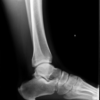
X-rays of the foot are important for the prevention of various injuries and diseases. With this diagnosis, both the presence of diseases and already running complications after severe injuries and bruises are determined. A person may feel unbearable pain when walking, this is a certain reason to take action on his part and perform radiography of the lower leg and foot, after which it is already possible to accurately identify the indicator of possible diseases. It is usually enough to take two pictures to establish an accurate diagnosis, but this is not always effective, because it may be necessary to conduct an x-ray at different angles to reveal a detailed picture of the disease.
This image shows soft tissues and bones of the foot comprising the Shin bone and tibia and fibula, constituting together the ankle, metatarsal bone, in the forefoot and phalanges. You may also be assigned to do oblique x-ray projection of the foot. The purpose of the image is the same as that of a direct projection image. A picture of the foot in a lateral projection in an upright position of the patient with an emphasis on the examined limb is made in order to detect flat feet.
X-ray refers to x-ray methods, since x-ray radiation that has passed through the part of the body under study is used to obtain images. In order to clearly understand what the foot x-ray shows and why you need to know the physical basics of radiography.

How do I get an x-ray?
Radiography of the foot is indicated in the following cases:
– You complain of pain in the foot area. Pain may occur when walking, after prolonged exercise, or at rest.
– Foot deformation. It is most often found in the area of the metatarsophalangeal joint and is characterized by a violation of the location of the phalanges of the thumb in relation to each other.
– This curvature leads to the appearance of a lump on the outside of the foot and becomes not only a cosmetic defect, but also brings a lot of unpleasant sensations and restrictions.
– Inflammation and infection of bones and joints – Arthritis, Arthrosis.
– Mechanical damage to the foot – blows, bruises, fractures.
– Platypodia.
– Congenital malformation of the foot.
Preparing for a x-ray of the foot
You do not need to prepare for an x-ray of the foot, but you must adhere to one condition-Before the x-ray, you must remove jewelry and accessories made of metal located in the study area. To protect you from excessive radiation, special lead aprons are applied to those parts of the body that are not subject to research.
The entire x-ray procedure of the foot can last about fifteen minutes: the immediate period of exposure to the rays does not exceed one second.
X-ray of foot and ankle
Indications for radiography of the foot and ankle:
– fracture;
– neoplasms;
– platypodia;
– osteoarthritis;
– rheumatoid arthritis;
– psoriatic arthritis;
– septic arthritis.
The foot is placed on a table or a special stool until the desired position is reached. If you take several pictures in different projections, our radiologists will periodically change the position of your foot. In addition, you may need to take a photo of your healthy leg if you need to make a comparison.
Right and left foot x-ray
Depending on your complaints and the intended diagnosis, the radiologist may need to perform a diagnostic imaging foot in different positions – it is used for a complete examination of the foot and its comparison with another. A common reason for this study is the presence of flat feet. This is a common disease that affects approximately 40-60% of the population, regardless of age and gender. Congenital or acquired deformity of the feet causes significant discomfort and leads to the development of severe disorders of the musculoskeletal system, joint inflammation, damage to the lower leg, varicose veins.
Effective treatment of pathology in a conservative way is possible only at the initial stage, so special attention should be paid to timely diagnosis, The most accurate method of which is radiography.
Ankle x-ray
For the procedure of this x-ray, there are certain prerequisites that are considered by surgeons, orthopedists and traumatologists. If you suspect gout, osteophytes, arthritis, osteoarthritis, or flat feet, or your lower leg starts to hurt a lot, you will be referred for an appropriate examination. On the other hand, the doctor may prescribe an x-ray of the ankle joint for other reasons, such as the presence of a tumor, degenerative change in the structure of the bone, or a suspected crack or fracture.
The method of radiography involves obtaining a two-dimensional image of the bone tissue of the studied area of the body. Structures with high density are clearly visible to the eye of a professional, who can use them to clarify the diagnosis and prescribe the necessary treatment. Soft tissue can also be detected on the x-ray of the ankle joint. The latter are indicated by a dark color, in contrast to the bones, through which the rays practically do not penetrate, and, consequently, the color of the bone structures in the image becomes white. You will be asked to extend your foot so that you can fix the front part of the limb and the ankle joint. In this case, the condition of the talus and tibia, the frontal segment of the ankle joint is evaluated. Tibia and fibula x-ray is performed. This type of x-ray examination is necessary if there is a suspicion of an injury in the ankle, which affected the tibia and fibula, or other elements of the forefoot.
In the image, the radiologist can identify all the pathologies of the joint, as well as determine how much the connective tissue was damaged. In this way, a number of dangerous conditions can be prevented, such as bone disposition, which can lead to loss of mobility and functionality of articular bone structures. Sometimes an x-ray of the ankle joint is not informative enough. In this situation, doctors prescribe a CT scan of the same joint.
The diagnostic imaging center is ready to provide you with the necessary help and give you some recommendations for subsequent treatment. Entrust the foot x-ray in Orlando, Florida only to the best, highly qualified radiologists, because we are able to see the cause of the problem and fix it.
How much do Foot X-ray’s cost
If you are interested in the question: “How much do X-ray’s cost in Orlando, FL?” – You can always give us a call to find out about your payment options.
*We accept health insurances.
*We accept patients with auto insurance after auto accidents as well as with letters of protection from attorney (LOP).
*We also accept self pay and care credit.
Costs can vary depending on the scan you need. Your insurance may cover the full cost of the scan or you might be responsible for part of the payment depending on your coverage.
Give us a call to find out more about your unique situation.
You can make an appointment today.
If you are looking for “X-ray of the foot near me”, then you have come to the right page. Our center is located near these locations:
College park, Apopka, Ocoee, Edgewood, Winter garden, Baldwin Park, Doctor Phillips, Millenia, Belle isle, Windermere, Pine castle, Altamonte, Pine hills, Metrowest, Downtown.

Other types of X-ray
Chest X-ray | Digital x-ray | Head and skull X-ray | Foot X-ray | Knee X-ray | Neck X-ray | Hand and Wrist X-ray | Shoulder X–ray | X-ray for children | X-ray Hip | X-ray Pelvis | X-ray Thoracic spine | X-Ray Orlando | Xray center
Our Google Reviews
Our Happy Clients


