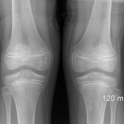
What is an x-ray of the knee joint?
One of the main reasons for disability of the active part of people is joint diseases. The popularization of complex sports leads to an increase in the number of injuries and orthopedic problems that occur at any age, which is why it is so important that their diagnosis is safe, accurate and as early as possible.
These requirements are fully met by the method of magnetic resonance imaging. Usually, an x-ray is done after consultation with an orthopedic doctor, who conducts special tests and makes a preliminary diagnosis.
To determine the cause of the joint malfunction, it is necessary to carefully study the condition of all tissues. Even the most minor damage to any of them can significantly affect the function of the entire lower limb.
Knee x-ray accurately visualizes all the degrees of cartilage change from edema to thinning, фflaking, and cracking. The condition of the sub-cartilaginous bone tissue, changes in the meniscus, inflammation of the synovial membrane, and so on are evaluated.
It is possible to calculate the total volume of the affected cartilage and altered bone sections, and assess the condition of the cruciate ligaments.

Knee x-ray
The knee joint is one of the largest and has a complex structure, which can be seen on x-rays. It plays an important role in the body, participates in a variety of movements and can withstand the weight of a person. Most calls to the traumatologist are associated with this part of the body. A large load can negatively affect the health of the knee joint. Knee x-ray gives an informative picture during the examination.
The knee joint includes the connection of the following units:
– bones, tibia and femur;
– patella, patella;
– cruciate ligaments;
– muscles;
– menisci;
– nerve endings and blood vessels.
All kinds of movements, in particular, turns, rotational movements, flexion and extension of the lower leg, are performed by the articulation of the knee joint.
Menisci – connecting elements. They distribute the load from the weight of your body, stabilize the entire joint. The menisci occupy a position between the cartilaginous surfaces of the main bones.
With the help of ligaments and muscles, two large bones are connected. To keep the patella in a fixed position, it is also connected by ligaments, the front of the structure is protected by the kneecap. There is a layer of cartilage to reduce friction and shock absorption.
Knee x-ray procedure
The x-ray image of the knee joint simultaneously includes the area of the knee, the area of the fibula, tibia, and femur. Equipment alone is not enough to see violations and make a diagnosis. Our specialist has a certain experience to know at what angle and from which side to do a knee joint x-ray each individual case, your foot is placed in a certain way, the projection is selected.
Special preparation before the x-ray is not required. There should be no clothing on the study area, if the knee is tied with a bandage, then it can be left. During the diagnosis, you should not move, our radiologist will definitely warn you about this. If you make a movement, the image may be blurred and it will be impossible to read the information.
What can you see on an x-ray of the knee?
X-rays of the knee joint will show clear boundaries of the joint ends, they look like a thin shadow, limited by the subchondral plate. If there are problems, the plate does not have smooth contours. The diagnostic image knee shows the following evaluation criteria:
– Speaking of the knee joint, the integrity of the tibia and femur is evaluated. If there is a fracture, its extent and location are described.
– In the absence of pathologies, the size of the joint gap is 5 mm. If its size is smaller, the joint is deformed or there is cartilage dystrophy.
– To assess the condition of the patella, the Cato index is calculated. Its index should be equal to the number 1.if the index is increased, then the ligaments are damaged. For the picture, the knee is bent at 300.
– Unfortunately, it is not possible to determine the bone density using this method. The image will show the swelling, structure and shape of the bones. In various degenerative diseases, fluid can be detected in the joint tissues.
– The fracture line is clearly visible in the image. If there is a crack, it is better to consider it not immediately, but after a few days, then it becomes more sharp. You can determine the dislocation by the following sign: the articular surfaces no longer correspond to each other.
– If the patellar tendon is damaged, the latter will occupy an unnatural position. The tear or sprain itself is not visible in the image, but the diagnosis can be made by the increased distance between the bones.
You can diagnose osteoarthritis if the cartilage is thin, the bone grows along the edges, and the bone is compacted near the cartilage. The joint gap in arthritis is enlarged, but one x-ray is not enough to make such a diagnosis. To understand that there is a cyst in the knee joint area, you can look at the light areas in the image. Osteophyte is also clearly visible, the shape of the bone changes due to the growth. If the bone in the picture is not very clear, translucent, then we may be talking about osteoporosis. This is caused by a lack of calcium in the study area. But the edges of the bone, on the contrary, are clearer compared to the norm.
The diagnostic imaging center provides high-quality x-ray services and offers its unique radiologists with special training and high qualifications. Make an appointment for a consultation right now and get a knee x-ray in Orlando, Florida according to all the accuracy of our radiologists.
How much do Knee X-ray’s cost
If you are interested in the question: “How much do X-ray’s cost in Orlando, FL?” – You can always give us a call to find out about your payment options.
*We accept health insurances.
*We accept patients with auto insurance after auto accidents as well as with letters of protection from attorney (LOP).
*We also accept self pay and care credit.
Costs can vary depending on the scan you need. Your insurance may cover the full cost of the scan or you might be responsible for part of the payment depending on your coverage.
Give us a call to find out more about your unique situation.
You can make an appointment today.
If you are looking for “X-ray Knee near me”, then you have come to the right page. Our center is located near these locations:
College park, Apopka, Ocoee, Edgewood, Winter garden, Baldwin Park, Doctor Phillips, Millenia, Belle isle, Windermere, Pine castle, Altamonte, Pine hills, Metrowest, Downtown.

Other types of X-ray
Chest X-ray | Digital x-ray | Head and skull X-ray | Foot X-ray | Knee X-ray | Neck X-ray | Hand and Wrist X-ray | Shoulder X–ray | X-ray for children | X-ray Hip | X-ray Pelvis | X-ray Thoracic spine | X-Ray Orlando | Xray center
Our Google Reviews
Our Happy Clients


